Diagnosing strokes with computed tomography (CT) scan
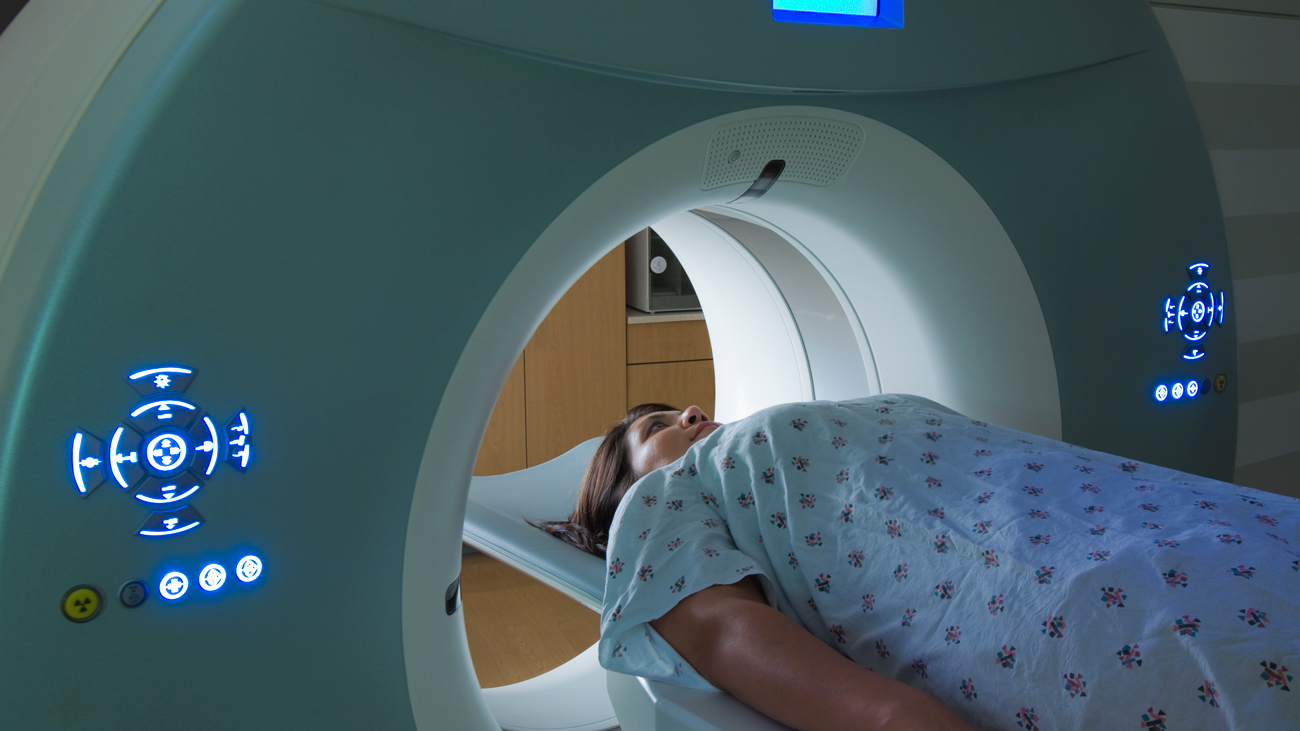
When doctors suspect that a patient might have had a stroke, tests are critical in making the correct diagnosis and applying the correct treatment as soon as possible. Often, the first imaging test for such a patient is a computed tomography (CT) scan. A CT scan uses common X-rays, but in a different way. The CT scanner takes multiple X-ray images a small distance apart, creating a cross-section view. The images look like “slices” of the patient’s head, giving doctors a three-dimensional look at the patient’s brain.
A brain CT scan can show bleeding in the brain or damage to the brain cells from a stroke. This test can also reveal other brain conditions that may be causing the patient’s symptoms.
Doctors often repeat CT scans during the treatment of a clot or blockage causing a stroke, to gauge the progress or effectiveness of the treatment.
Sometimes doctors will repeat CT scans after giving the patient an intravenous dye or “contrast agent.” This produces clearer images of the blood vessels inside the brain, usually not visible in CT images. This can identify which blood vessels are blocked and can help determine how to treat the patient, or to show if treatment is working to break up clots and unblock blood vessels.
St. Charles Bend in Central Oregon offers a wide range of services to prevent, diagnose and treat strokes.